
Dermatology, like other areas of medicine, has experienced and continues to undergo enormous changes in both technologies and in medicine and procedures. Dermatologists and other experts involved in skin care, attribute much of this transformation to fundamental improvements in medicine. With today’s innovative advancements in skin care, people can now get relief from age-old problems like acne and scarring. There’s a revolution going on in the area of treating aging skin.
Many dermatologists are now using vitamins and minerals directly on the skin. Laser treatments have proven quite effective as well. Regardless of whether you’re dealing with stubborn pockets of fat or varicose veins, there’s a new treatment for it.
The advent of more advanced imaging capabilities, telecommunication systems, and methods of information transfer have significantly changed how dermatologists examine patients. They can now perform an examination, develop a diagnosis, and outline a treatment plan using software specifically developed for this field.
Technology is in a constant state of growth and the transformation it brings to every industry is enormous. After canvassing dermatologists and their colleagues in the skin care industry, we have put together a wealth of information on some of the most effective ways in which these experts have employed various forms of technology in their work. Read along.
Clinical Photography in the practice of dermatology
Dermatologists have used photography for decades as a conventional means for documenting skin conditions. Photography comes in handy as an adjunct to the treatment of various dermatologic conditions in both research and clinical applications.
This approach to capturing and documenting patient improvement has been furthered by the emergence of low-cost digital imaging systems that are considerably easier to use. There have been improvements in the imaging applications as well. Many skin care professionals have used these new approaches but others have concerns when it comes to using digital imaging technologies to document dermatological information. These concerns are:
- Controlling the lighting to provide a clear record of the skin condition of patients.
- Maintaining consistency over time to keep an accurate track of clinical endpoints for meaningful comparisons.
Ideally, you’ll want to control the image capturing for quality, making sure that the images can be reproduced and compared with ease. This is the key to the successful use of clinical photography in dermatology. It means controlling factors that typically influence image quality such as:
- Lighting (the lighting has to be right)
- Distance (image must be taken from a close distance to produce optimum image resolution)
- Color reproduction (should be realistic)
- Focus (sharp is preferred)
- Distracting elements (on the background/avoid them as much as possible)
- Points of reference (the inclusion of a chosen point of reference is helpful)
Note: Photos will be most suitable for purposes of comparison when taken under identical conditions and at the same angle, using the same center of focus each time.
With increased accessibility to improved digital imaging systems, practitioners have been able to capture good-quality photographs of affected areas for diagnosis. The fact that digital imaging equipment has become more universal among today’s consumers has contributed to improved functionality in the medical industry.
There are numerous ways in which dermatologists have used clinical photography in their work over the years. Here, we look at some of the best ways in which these technologies have been used to capture and document a patient’s skin information.
Using photo analysis software and 3D imaging tool within dermatology clinics
Various dermatology clinics have turned to 3D imaging tools to photo document cutaneous disorders largely due to the visible nature of the skin. This has been an increasingly more convenient and cheaper undertaking given the emerging developments in imaging technology.
Ongoing technological advances have constantly created more and more opportunities for photo taking and use in dermatology clinics. There is widespread availability of quality low-cost digital cameras for clinicians to use in capturing images for patient records. This has enabled dermatologists to easily rely on standard photography as a useful technology in their practice; both for medical and cosmetic applications.
Physicians have found photos to be extremely helpful in documenting their patients’ short-term or even long-term clinical history; largely because patients often forget how they looked prior to therapy. Photos, therefore, become handy as patient management or education tools.
Clinical trials: using software to analyze images
While photo documentation has been at the center of clinical trials for several dermatologic indications or diseases for many years, developments in technology have improved it even further, making it more essential in these clinical trials.
The most successful use of documented photographs for clinical and cosmetic complaints have involved using software to analyze images and provide objective and more accurate assessments of various patients’ therapeutic responses. These include the use of both two-dimensional (2D) and three-dimensional (3D) imaging systems developed for clinical trials. These methods have been far more dependable than any human reviewers.
Together with specially designed imaging software, these systems have been employed to offer accurate measurements of objective criteria including lesion sizes and lesion counts.
Further, with 3D imaging technology, dermatologists have been able to get better measures of essential dermatologic parameters such as wrinkle depth. Without this kind of technology, experts had to previously rely on less accurate assessment methods like comparing before and after images of the skin in focus. This kind of approximation left much room for error. However, with the advent of 3D imaging technology, dermatologists have been able to obtain more accurate measurements of essential factors including the height, width, and even changes in lesion volume which can provide a more accurate evaluation of the effect of therapy on different lesions.
How 3D imaging technology works
It is easier to comprehend the working of three-dimensional technology if looked at from the perspective of the working of the human eye. Our eyes perceive depth (3D) since each eye, located some inches apart, views the same object from an angle slightly different from the other. The brain then merges this data to create a 3-dimensional representation of what we are looking at.
The three-dimensional imaging system works in basically the same fashion. It utilizes a compact stereovision camera to capture two images that differ from each other by some degrees, for every photo taken. Like the human brain, the system then combines the two images (taken at different angles) and forms a single composite three-dimensional image of the object.
Once clinicians have these life-like reconstructions the system has created, they turn to 3D analysis software to show a variation from baseline. The software can also be simulated to help indicate or predict a response expected from treatment.
Storage and retrieval of 3D images
Documentation of 3D images has been successful to a large extent because of the software for image storage and management that makes up part of the 3D imaging technology.
Dermatologists often attend to multiple patients and this means they may have thousands of patient photos stored in different places (libraries, disks, computers) nearly at every point in time.
Retrieving a patient’s image from such a mammoth repository can be tedious if you don’t have a mechanism in place to simplify the process. Not to mention that there is usually no standard formatting designed for file naming and storage in the dermatology practice. The common habit has been to tag a file, along with the name of the patient and presentation date.
Thanks to advanced image storage and retrieval systems that centralize and standardize image storage, clinicians have been able to link more patient images with their (patients’) records.
Such state-of-the-art software offers a more standardized and effective means of recording and searching for a patient’s image(s) when required. Further, it has been easier for practitioners to conduct side-by-side comparisons on-screen with this kind of technology.
New technologies that have been incorporated into clinical photography
Imaging systems and software used in clinical trials have seen major changes for the better. Some of the new capabilities that have since been incorporated into these technologies include the use of precisely angled light pointers on both sides of the image processing lens, enabling the user to pinpoint the specific object to be snapped (such as anatomic site or lesion).
This is a significant addition since it enables some of the most essential requirements for accurate imaging to be achieved. For instance, the technology makes it easier to get the exact image distance and camera settings that are needed to achieve consistency in images over the entire period of treatment, even when multiple users are involved in taking the photographs over that extended period of time.
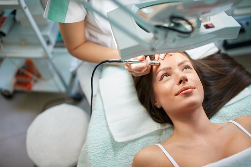
Laser technologies for skin imaging and treatment
Other than traditional clinical photography and regular 3D imaging technology, laser technology has made even greater contributions in helping dermatologists achieve efficiency in both cosmetic and clinical therapies.
There have been state-of-the-art options that enable these clinicians to view far more details that cannot be seen any other way. Further, the use of laser appliances has enabled dermatologists to produce incredible, microscopic skin images and analyze data in a more in-depth manner.
Laser technology for skin diagnosis
To analyze the patient’s skin, clinicians have always had to take captured images to the lab for special analysis. Over the recent years, this has been rendered unnecessary in most cases. With the recent improvements of laser technology and its incorporation in the field of dermatology, it has become less common for dermatologists to have to peer at glass slides on a microscope.
A good number of clinicians have used dermatoscopy, a technique that involves using a cutting-edge, hand-held microscope to examine the skin. The device is built for studying the complex surface of the skin and captured images or recorded videos as needed. This process requires absolutely no glass slides under the lens of the microscope.
Dermatoscopy also allows dermatologists to look beneath the skin by use of a special optical imaging technique involving the use of a confocal laser scanning microscope. This technique reveals at a cellular level, the very root of a broad range of skin conditions.
Thanks to the confocal laser scanning microscopy, patients with various skin conditions have enjoyed noninvasive procedures enabling them better results with faster recovery times. The technology is also available with other equipment used for mole mapping or full body imaging.
These developments have been responsible for accurate and real-time imaging that help prevent disease through early detection. There have also been significant improvements in understanding the nature of skin itself.
Noninvasive treatment options
The application of laser technology does not just end with diagnosis; rather, it further helps in the subsequent stages that involve providing solutions to the patient.
Noninvasive treatment is one of the areas in which laser applications come in. This form of treatment does not involve cutting the skin. It has transformed medicine in significant ways, particularly in the field of dermatology.
Advancements in laser technology have made them far more accurate with precise targeting that leaves healthy skin completely untouched. For instance, fractional lasers have been used to deliver numerous tiny laser beams in a grid pattern – treating only the affected skin cells and leaving alone the surrounding cells as well as the cells in between.
These laser options have shown significant efficacy in treating skin conditions with negligible damage to the skin. They are a much less damaging alternative compared to the traditional use of the scalpel. The laser is also more desirable especially for treating delicate areas of the skin.
With the noninvasive nature of laser treatments, patients of dermatologic procedures have registered some of the shortest recovery times in the history of medicine. The outcomes have been great for medical and for cosmetic needs alike.
In many cases, dermatologists have applied the use of the same lasers used in treating lesions or skin cancers to cosmetic applications such as treatment of skin aging, sun damage or even removing undesirable pigmentation, such as from acne or scars.
At-home laser hair-removal techniques
Laser use is not just limited to the confines of dermatology clinics. Over the years, innovators have worked hand-in-hand with leading dermatologists to create some very effective at-home laser appliances for hair removal.
Some people, mostly females, tend to prefer smoother and clearer legs, considering hair to be unwelcome on the skin surface. For such people, there have been impressive results from the use of appliances like Tria Beauty’s Laser Hair Removal System and SensEpil from Silk’n.
With laser technology, what we see is a unified approach to skin treatment for both clinical and cosmetic needs. A lot of dermatology clinics have invested in proven technologies and equipment that work so that patients have enjoyed therapy without the need for surgery.
Keeping up with the demands associated with growing medical literature
Like other areas of medicine, dermatology is constantly evolving. This means that there is increasingly more medical literature for the dermatologist to utilize in order to deliver improved healthcare services.
To keep up, dermatologists have resorted to the use of information technology tools to record, organize, manage and/or recall every ounce of essential information. They are turning to electronic health record systems and the cloud for easier access to needed data in its electronic format. Personal computers, mobile phones, tablets, and other related information have become indispensable resources.
In so doing, they have automated a huge bulk of the information management work and freed up large amounts of time to use in developing better treatment methods for their patients.
There has been positive feedback from both patients of cosmetic dermatologic procedures and their physicians. This points to the positive impact that the use of information communication technologies has had on this industry.
In other words, these technologies, with their multimedia capabilities including drawing tools and videos, have been responsible for improved clinical efficacy. Patient satisfaction has increased, along with overall patient experience.
Effective sharing of information via Internet-based dermatology resources
Information communication technologies have not been limited just to dermatology clinics and consultation rooms. Dermatologists and other clinicians have turned to web-based apps and systems to transfer or share information in a social and collaborative manner – effectively transforming the whole arena of access to data and physician-patient communications in general.
Dermatologists have moved a step further to create information resources such as blogs and vlogs, as well as eBooks and other publications, making them electronically available for patients and colleagues to access. These resources are being used for educational purposes by many experts.
Patients have often already checked out all the treatment options online, so they have a better idea of what to expect from therapy – effectively reducing consultation time and improving both patient experience and treatment outcomes.
Because of this growing use of internet-based dermatology resources, physicians can now easily access learning and reference materials online. Lots of educational support resources are also available for patients online, which have helped improve dermatology and continue to facilitate the growth of the practice.
Last words
The last few decades have seen tremendous technological advances and innovations in dermatology and dermatologic procedures or surgery. These have slowly but progressively changed how dermatologists interact with patients and fellow physicians, as well as the way they practice medicine overall.
With new tools and approaches to skin care and related therapies, dermatologists have had some of the finest tools and equipment at their disposal. These professionals have made great use of these technologies to bring improved care to the patient. Based on this record, we can expect to see nothing but a stream of constantly-improved healthcare from dermatologists going forward.